Exam of the Internal Reproductive Tract: Palpation & Ultrasound
Rectal palpation and ultrasound should be performed only by trained personnel since there is a chance of serious injury/death to the mare from rectal tears. In order to protect the mare, appropriate restraint should be used (stocks, anaesthesia).
Rectal palpation: allows the veterinarian to examine the cervix,uterus and ovaries.
- Cervix- size and tone indicates progesterone levels
- Uterus- tone & size of horns (indicate pregnancy, stage of cycle), pregnancy, fluid
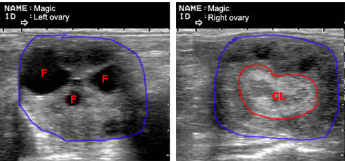
- Ovaries- size & activity (follicles, early CH, CLs > 5 days)
Ultrasound: shows more detail than can be determined by rectal palpation.
- Uterus- edema (intra-tissue fluid indicative of estrus), small pockets of fluid, characteristics of the fluid, endometrial cysts (size and location recorded for future reference), early pregnancy, twins.
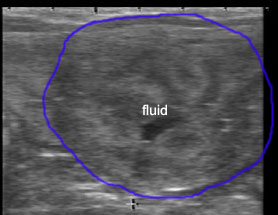
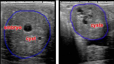
Estrual uterus shows edema and clear fluid in lumen (normal). Embryo and cysts in the uterus. It is important to know the size and location of cysts before pregnancy since a cyst can be mistaken for an early pregnancy. (see Hysteroscopy image of a cyst)
.
- Ovaries- size and shape of follicles, CL, CH, hemorrhagic follicles, anovulatory follicles, granulosa cell tumors, hematomas