
Uterine Culture, Cytology & Biopsy, Hysteroscopy
Many responsible stallion owners require a "clean uterine culture" from a mare prior to breeding her live cover or shipping semen. The results of an endometrial swab culture alone are not a reliable indicator of uterine pathogenicity since it may reflect contamination of the sample. A cytology smear of the sample can be taken in conjunction with the culture to identify mares with active infection (increased inflammatory cells: neutrophils, lymphocytes, plasma cells, eosinophils, macrophages, and mast cells). Immediate results on the farm are possible from cytology and can indicate treatment although culture is required to determine the specific organisms present and treatment options (specific antibiotics).
- Culture & cytology of the uterus should be considered as a single procedure
- The best time to perform the procedure is during (late) estrus because uterine flora decreases with increases in estrogen
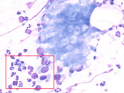
Low power: Abnormal uterine cytology 
High power: Inflammatory cells and bacteria
Endometrial Biopsy
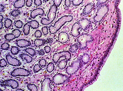
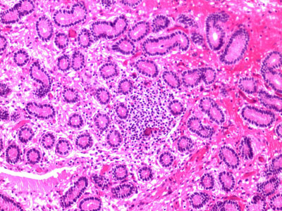
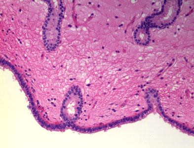
An endometrial biopsy takes a piece of the uterine surface to detect inflammation or degeneration. Scar tissue also indicates a chronic irritation/infection. The endometrial biopsy is usually the last step in the breeding soundness examination and is indicated in pre-purchase exams, barren mares, mares with past infertility, abortions, pyometritis, and mares requiring reproductive surgery. When to perform a biopsy duing the cycle is a matter of choice and availability:
- estrus: the cervix is open and more easily penetrated and the mare is more resistant to endometritis
- diestrus: the endometrial glands are more active and may give a better picture of their function
The biopsy is assigned to a category based on pathological evaluation.
| Category | Pathology | Foaling rate |
Factors effecting foaling rate |
| Category I | normal, pathologic changes slight and sparsely scattered | 80-90% |
overall management |
| Category IIA | mild changes | 50-80% |
overall management, endometrial changes |
| Category IIB | moderate changes | 10-50% |
overall management, endometrial changes |
| Category III | severe changes: widespread inflammation, scarring, atrophy of glands in the physiological breeding season | 0-10% |
severe changes in the endometrium |
Histological examples of biopsy:
|
 |
|
 |
|
 |
Hysteroscopy
This procedure is performed at a well equipped veterinary clinic to aid in the diagnosis of the causes of infertility or to help in the placement of semen when only a small number of sperm are inseminated. A small endoscope is passed into through the cervix into the uterus to visualize the cervix, uterus and uterine horns in 3 dimensions.
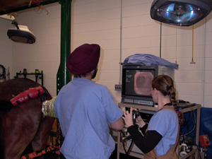 |
Hysteroscopy setup (UW -Madison Veterinary Hospital). "Real time", 3 dimensional pictures of the uterus can be viewed as the camera is manipulated in the uterus. (Pictures provided by Dr. Harry Momont) |
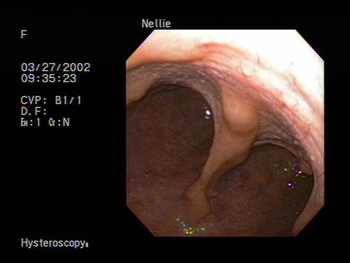 |
Uterine bifurcation (where the uterine horns separate from the uterine body) |
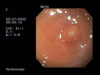 |
Oviductal Papilla: at the tip of the uterine horn where the oviduct begins. This is the site of semen placement when inseminating with low concentration of semen to increase the chance of pregnancy. |
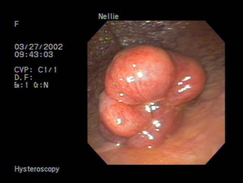 |
Uterine cysts (see the ultrasound image of cysts) |
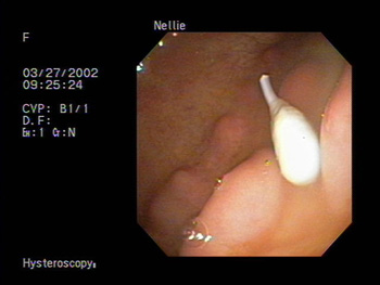 |
Uterine "foreign body" |
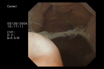 |
Fungal growth in uterus. |
You are now done with this lab!