
Pregnancy Detection
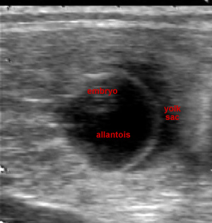
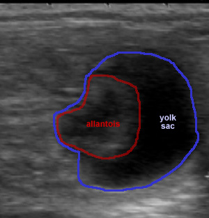
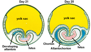
The following is a scan of a day 23 pregnancy and then a diagram from the lecture material on a day 21 and 25 embryo. At this point, all 4 parts of the placenta have begun to develop and consist of the yolk sac, amnion, allantois and chorion. These are actually a series of sacs that develop around the embryo. Only the amnion, the inner most sac is not labeled in the diagrams. Be sure you can identify the structures both in the ultrasound images and the diagram. The most prominant features at this age that are visible on ultrasound are the allantois and yolk sac.
| Mare: Lady 3/23/06 - Day 23 Characteristics:
|
|
 |
 |
|
|


Pregnancy