
Pregnancy Detection
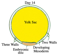
The following is a scan of a potential day 14 pregnancy. View the scan and answer the questions. Keep track as to when you are in the right and left uterine horns. Remember that this is same mare from which the pre-pregnancy ultrasound image and cyst map was obtained. At the bottom is a diagram of an embryo of this age. The embryonic disk are the cells that will develop into the fetus. The first of 4 placental membranes has developed and is called the yolk sac. The yolk sac is the fluid you are seeing on ultrasound at this stage of development. the embryonic disk is not visible on ultrasound at this time.
| Mare: Lady 3/14/06, c. checura - Day 14
|
How many embryonic vesicles are seen in this mare?
Why is one fluid filled structure smaller?
|


Pregnancy