Animal Science 434 - 1/26/98
A. The Testes. These primary organs have the dual function of producing sperm cells, and also the male hormone, testosterone. The testis is enclosed in the tunica albuginea surrounded by another tough tunic, the tunica vaginalis. The seminiferous tubules are the site of sperm formation. These in turn empty into collecting ducts, the rete testis, lined with cuboidal epithelium. The supporting connective tissue joins centrally forming a fibrous cord, the mediastinum testis.
B. The secondary sex organs are the ducts and tubes which convey the sperm cells out of the testes and eventually out of the body. They are listed in order in which sperm pass through them.
C. The accessory sex glands
D. Protective, supporting, and other structures
The general location of the different parts of the reproductive tract
of the bull is shown in the following diagram.
While it is possible to show only 1 testis clearly in this diagram there are two testes, and two sets of ducts carrying the sperm to the urethra. The urethra is a single duct, which also carries urine from the bladder.
The testes are oval-shaped organs 4 to 5 inches in length, 2-1/2 to 3 inches in diameter, with the long axis being vertical. Each testis weighs 10 to 12 ounces in a mature bull. They lie outside the body cavity in a pouch of skin called the scrotum. An important purpose of the scrotum is to provide the testes with an environment which is a few degrees (2-8°C) cooler than body temperature. This cooler temperature is necessary for the formation of spermatozoa. Failure of the testes to descend from the abdomen into the scrotum, associated normally with shortening of the gubernaculum and intra-abdominal pressure, results in a condition known as cryptorchidism. This will cause sterility if both testes fail to descend (bilateral cryptorchidism). Unilateral cryptorchid animals may be fertile, but it is thought that this condition may be inherited, and breeding males possessing this trait should be avoided.
For many years the external cremaster muscle within the spermatic cord has been thought of as the principle thermoregulator of the scrotum, drawing the testes close to the abdomen when cold and relaxing when warm. However, the tunica dartos muscle at the bottom of the scrotum also responds to temperature changes and probably plays a major role in temperature regulation of the testes. It has been shown that this latter muscle is sensitive to temperature changes only in the presence of the male hormone, testosterone. Blood flowing to the testis is cooled by adjacent venous return in a convoluted complex of vessels called the pampiniform plexus, located just dorsal to the testes.
The testes are partially supported by the spermatic cord, which runs from the abdomen and is attached to the testes in the scrotum. This band of tissue, carrying the ductus deferens, blood vessels, nerves and muscles associated with the testis, may be 8 to 10 inches or more in length.
The many convoluted seminiferous tubules in the testis in which the spermatozoa are formed finally straighten and join to form the rete testis. Arising from the rete testis are 12 or more out-going ducts, the vas efferentia, which emerge from the testis and enter the epididymis. The epididymis is a single large tortuous tubule lying on the surface of the testis. Its purpose is to collect and store the sperm while the latter undergo a ripening process. The different parts of the epididymis are referred to as the head, the body, and the tail (caput, corpus and cauda). It is the tail of the epididymis that the majority of sperm are stored. The relationships of these structures are shown in Fig. 2.

From the epididymis, the spermatozoa are carried into the vas deferens, the tube which carries the testicular products to the urethra of the penis. The vas deferens are very small in diameter, having a cartilaginous cord-like appearance, and each forms a saccular ampulla, 4 to 5 inches long and 1/2 inch in diameter, near the junction with the pelvic urethra. The two ampullae empty into the urethra. The urethra serves as the common passage for urine, and upon ejaculation for semen.
The organ of copulation is the penis. In the adult bull it is about 3 feet in length and about 1 inch in diameter. There is very little erectile tissue present except in the root. Even in the relaxed state the penis is very dense and firm. Behind the scrotum it forms an S-shaped curve, the sigmoid flexure. During erection this flexure is straightened out, thus increasing the length of the organ. Also, at the time of erection the bulbospongiosus and ischiocavernosus muscles at the root of the penis contract and assist in causing an erection and in ejecting the semen. The retractor penis muscle assists in withdrawing the penis into the sheath after copulation. The sheath is long and narrow. The preputial opening is a short distance behind the navel and is usually surrounded by long hairs.
The accessory glands associated with the reproductive tract, secrete a large part of the semen. In most species, these are the seminal vesicles, the prostate, and the bulbo-urethal or Cowper's glands (see Fig. 1). The paired seminal vesicles are tortuous lobulated glands, which produce a large volume of fluid which may flush the urethra and act as a vehicle for sperm transport. This secretion also includes fructose. The prostate is a compound gland lying over the urethra at the neck of the bladder. It produces a complex secretion, which stimulates sperm activity. The paired Cowper's glands lie below the prostate on either side of the urethra, and produce a viscid mucus-like lubricating substance. The secretions of all of the accessory glands plus the spermatozoa and the secretions of the testes and tubes leading from it make up the semen. These secretions aid in the nutrition of the sperm and provide some buffering capacity.
Cross-Section of the Testis
If one takes thin slices from the testis and examines them with a microscope at high magnification the testis is seen to be full of tiny tubules, the seminiferous tubules. These tightly coiled tubules, thousands of yards in total length, are lined with germinal epithelium, from which the sperm cells are eventually formed. In between the tubules is found the interstitial tissue. This tissue produces the male hormone, testosterone. Also, this tissue gives some support to the testis. The cut seminiferous tubules and interstitial tissue are shown diagrammatically in Fig. 3.

Reproductive System of the Boar
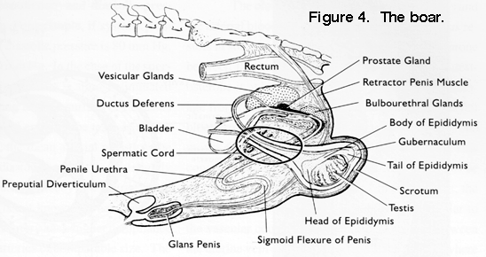
The preceding information on the bull applies in a general way to the boar. The names of the different parts of the reproductive system are the same. However, the relative sizes and arrangements of the various parts may differ from the bull as shown in Fig. 4. The testes of the boar are relatively large. They are suspended in an inverted position (compared to the bull and ram) with the tail of the epididymis uppermost. Particularly striking are the large Cowper's glands. These are the source of the gelatinous material in boar semen. Also, note the rotation of the testis relative to the vertical position in the bull. The penis has no true glans at the tip. The preputial pouch is pronounced in the boar. This contains some residual urine and decomposing epithelial cells which contribute to the unpleasant odor which may permeate boar meat.
Figure 4. The Boar.

Reproductive System of the Horse
The reproductive system of the stallion is shown in Fig. 5. Several differences from the bull and boar are distinctive. In the relaxed state the testes are nearly horizontal. They vary greatly in size depending upon the breed. The body of the epididymis is large. The penis is a vascular-muscular type and so enlarges greatly during erection. Considerable smegma accumulates in the preputial cavity and the penis should be washed before collecting semen or before copulation. There is no sigmoid flexure.
The Cowper's glands (bulbo-urethral glands) are about 1-2 inches in diameter and can be palpated rectally, in contrast with the bull. The seminal vesicles and prostate also can be palpated rectally near the junction of the bladder and the urethra.
Figure 5. The Stallion.
